Is An Ultrasound Safe
Yes!
Ultrasound is considered one of the safest ways to evaluate tissues and organs in your body especially compared to other testing modalities such as CT scans.
Ultrasounds do not contain radiation but instead use sound waves that bounce back off of your tissues at different rates and frequencies.
These sound waves vary in intensity and the amount of energy that they produce.
These variations result in images that Doctors can use to diagnose certain conditions.
How Can A Thyroid Ultrasound Help With Diagnosis
An ultrasound can give your doctor a lot of valuable information, such as:
- if a growth is fluid-filled or solid
- the number of growths
- where the growths are located
- whether a growth has distinct boundaries
- blood flow to the growth
Ultrasounds can also detect a goiter, a swelling of the thyroid gland.
Who Interprets The Results And How Do I Get Them
A radiologist, a doctor trained to supervise and interpret radiology exams, will analyze the images. The radiologist will send a signed report to the doctor who requested the exam. Your doctor will then share the results with you. In some cases, the radiologist may discuss results with you after the exam.
You may need a follow-up exam. If so, your doctor will explain why. Sometimes a follow-up exam further evaluates a potential issue with more views or a special imaging technique. It may also see if there has been any change in an issue over time. Follow-up exams are often the best way to see if treatment is working or if a problem needs attention.
Recommended Reading: Best Selenium Supplement For Thyroid
How The Test Is Performed
Ultrasound is a painless method that uses sound waves to create images of the inside of the body. The test is often done in the ultrasound or radiology department. It also can be done in a clinic.
The test is done in this way:
- You lie down with your neck on a pillow or other soft support. Your neck is stretched slightly.
- The ultrasound technician applies a water-based gel on your neck to help transmit the sound waves.
- Next, the technician moves a wand, called a transducer, back and forth on the skin of your neck. The transducer gives off sound waves. The sound waves go through your body and bounce off the area being studied . A computer looks at the pattern that the sound waves create when bouncing back, and creates an image from them.
What Is Thyroid Ultrasound Cpt Code

CPT code 2023 is used to report a diagnostic thyroid ultrasound. This code involves the use of real-time ultrasonography to image the thyroid gland and may also include Doppler imaging to assess blood flow within the gland.
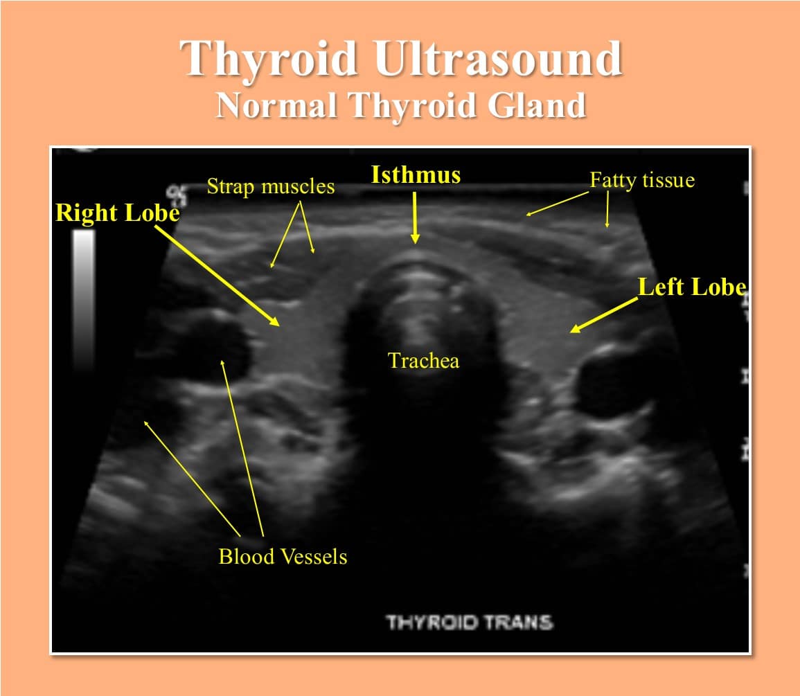
normal thyroid ultrasound
1. Normal thyroid ultrasound images usually show a well-defined gland with smooth borders and no evidence of nodules, cysts, or other abnormalities. Echogenicity is usually uniform throughout the gland.
2. In some cases, small areas of calcification may be seen within the gland, but this finding is not considered abnormal. The presence of a dominant follicle is also considered normal and should not be a cause for concern.
3. If no lumps or other abnormalities are identified on thyroid ultrasound, further investigation with additional imaging or biopsy may rule out thyroid cancer or other malignancy.
Read Also: What Can Help An Underactive Thyroid
Why The Test Is Performed
A thyroid ultrasound is usually done when physical exam shows any of these findings:
- You have a growth on your thyroid gland, called a thyroid nodule.
- The thyroid feels big or irregular, called a goiter.
- You have abnormal lymph nodes near your thyroid.
Ultrasound is also often used to guide the needle in biopsies of:
- Thyroid nodules or the thyroid gland — In this test, a needle draws out a small amount of tissue from the nodule or thyroid gland. This is a test to diagnose thyroid disease or thyroid cancer.
- The parathyroid gland.
- Lymph nodes in the area of the thyroid.
How To Prepare For An Ultrasound
Your ultrasound will probably be performed in a hospital. A growing number of outpatient facilities can also perform ultrasounds.
Before the test, remove necklaces and other accessories that can block your throat. When you arrive, youll be asked to remove your shirt and lie on your back.
Your doctor may suggest injecting contrast agents into your bloodstream to improve the quality of the ultrasound images. This is usually done with a quick injection using a needle filled with materials such as Lumason or Levovist, which are made of gas filled with tiny bubbles.
Recommended Reading: How Long Does Thyroid Eye Disease Last
How Should I Prepare
Once the imaging is complete, the technologist will wipe off the clear ultrasound gel from your skin. Any portions that remain will dry quickly. The ultrasound gel does not usually stain or discolor clothing.
After an ultrasound exam, you should be able to resume your normal activities immediately.
A radiologist, a doctor trained to supervise and interpret radiology exams, will analyze the images. The radiologist will send a signed report to the doctor who requested the exam. Your doctor will then share the results with you. In some cases, the radiologist may discuss results with you after the exam.
Ultrasound exams are very sensitive to motion, and an active or crying child can prolong the examination process. To ensure a smooth experience, it often helps to explain the procedure to the child prior to the exam. Bring books, small toys, music, or games to help distract the child and make the time pass quickly. The exam room may have a television. Feel free to ask for your child’s favorite channel.
What Happens During The Procedure
Ultrasound imaging uses sound waves to create an image of the thyroid, any lumps and/or nodules. It is the same technology used to perform antenatal scans during pregnancy. You will not be exposed to any radiation during the process.
It is important to remember that the doctors carrying out your ultrasound are there to answer any questions you might have at any time and to ensure you remain as comfortable as possible. If you have any questions, this is the time to ask them.
You may be required to wear a hospital gown for the ultrasound scan.
Once in the treatment room, you will be asked to lie on a bed with a pillow to support your shoulders. You will be asked to tilt your neck backwards to allow the best access to the area. A cool lubricant gel may be applied to the surface of the skin to assist the movement of the probe. The ultrasound probe will make contact with the skin and be moved around the neck. This will form images on the monitor allowing the sonographer or radiologist to assess and measure what the ultrasound has enabled them to see. Usually, these images will then be passed to your specialist before the results are discussed with you.
You May Like: How Fast Does Anaplastic Thyroid Cancer Grow
Ultrasound Of The Thyroid Gland
All iLive content is medically reviewed or fact checked to ensure as much factual accuracy as possible.
We have strict sourcing guidelines and only link to reputable media sites, academic research institutions and, whenever possible, medically peer reviewed studies. Note that the numbers in parentheses are clickable links to these studies.
If you feel that any of our content is inaccurate, out-of-date, or otherwise questionable, please select it and press Ctrl + Enter.
Where to make ultrasound of the thyroid gland and why it is necessary to regularly undergo preventive examinations of this body? The thyroid gland is a part of the endocrine system, the disease or disruption of its functioning negatively affects the work of the whole organism. Ultrasound diagnosis allows to identify in time the foci of pathologies and to conduct treatment.
Uses For A Thyroid Ultrasound
A thyroid ultrasound may be ordered if a thyroid function test is abnormal or if you doctor feels a growth on your thyroid while examining your neck. An ultrasound can also check an underactive or overactive thyroid gland.
You may receive a thyroid ultrasound as part of an overall physical exam. Ultrasounds can provide high-resolution images of your organs that can help your doctor better understand your general health. Your doctor may also order an ultrasound if they notice any abnormal swelling, pain, or infections so that they can uncover any underlying conditions that might be causing these symptoms.
Ultrasounds may also be used if your doctor needs to take a biopsy of your thyroid or surrounding tissues to test for any existing conditions.
Recommended Reading: Stop The Thyroid Madness Com
What Is Thyroid Ultrasound Images
There are several different types of thyroid ultrasound images, and each can provide valuable information about the health of your thyroid gland. A thyroid ultrasound images can help identify benign or malignant nodules, as well as assess the size and function of your thyroid gland. thyroid ultrasound images are usually quick and painless, and can be done on an outpatient basis.
Where Is It Usually Performed

An ultrasound scan is usually carried out at a hospital, usually in the endocrinology Endocrinology “the branch of physiology and medicine concerned with endocrine glands and hormones” or radiology Radiology “the science that uses x-rays or nuclear radiation to diagnose and sometimes also treat diseases within the body” department. It is a non-invasive procedure that does not require any anaesthetic Anaesthetic “a substance that causes lack of feeling or awareness, dulling pain to permit surgery and other painful procedures” .
Read Also: Selenium For Thyroid Eye Disease
How It Is Done
This test is done in an ultrasound room in a doctor’s office or hospital.
You may be asked to undress above the waist. And you may drape a paper or cloth covering around your shoulders. Remove all jewelry from your head and around your neck.
You will lie on your back on a high table with your neck stretched out. You’ll have a pillow under your shoulders. Gel will be spread on your neck. This helps the sound waves pass through better. A small water-filled bag or gelatin sponge might be placed over your throat. This also helps to conduct the sound waves. The transducer will be pressed against your neck . Then it will be moved back and forth over your neck. A picture of your thyroid gland and the tissue around it can be seen on a video screen. You may be asked to turn your head away from the side being scanned so your jawbone is out of the way.
You may be asked to wait until the radiologist has reviewed the information. He or she may want to do more ultrasound views of your neck.
What An Ultrasound Tells You
The ultrasound gives you information predominately about the shape, size, and texture of your thyroid gland.
All of these variables can help you to determine what is happening in your thyroid gland.
For instance:
Maybe youve felt a lump or a bump on your thyroid gland. An ultrasound will help you to determine if this lump is something serious or something benign .
Maybe youve already had an ultrasound and you are following up on a thyroid nodule to see if its grown over time. If youve been diagnosed with a thyroid nodule before then you will probably need frequent testing and monitoring.
Maybe youve been diagnosed with Hashimotos Thyroiditis and your Doctor wants to evaluate for inflammation in your gland. An ultrasound can check to see if you have inflammation throughout your gland which may affect how you feel.
Maybe you are in the postpartum period and your Doctor suspects you may have postpartum thyroiditis. An ultrasound can help determine if you have inflammation in your gland which may cause symptoms such as depression or weight gain in the postpartum period.
A thyroid ultrasound will also give you information about the characteristics of thyroid nodules which can give you an idea as to how likely they are to be cancerous.
The good news is that because getting a thyroid ultrasound is so easy the treatment for thyroid cancer is effective up to 97% of the time.
Recommended Reading: How Can Thyroid Problems Affect You
What Are Some Common Uses Of The Procedure
An ultrasound of the thyroid is typically used:
- to determine if a lump in the neck is arising from the thyroid or an adjacent structure
- to analyze the appearance of thyroid nodules and determine if they are the more common benign nodule or if the nodule has features that require a biopsy. If biopsy is required, ultrasound-guided fine needle aspiration can help improve accuracy of the biopsy.
- to look for additional nodules in patients with one or more nodules felt on physical exam
- to see if a thyroid nodule has substantially grown over time
Because ultrasound provides real-time images, doctors may use it to guide procedures, including needle biopsies. Biopsies use needles to extract tissue samples for lab testing. Doctors also use ultrasound to guide insertion of a catheter or other drainage device. This helps assure safe and accurate placement.
How Does The Procedure Work
Ultrasound imaging uses the same principles as the sonar that bats, ships, and fishermen use. When a sound wave strikes an object, it bounces back or echoes. By measuring these echo waves, it is possible to determine how far away the object is as well as its size, shape, and consistency. This includes whether the object is solid or filled with fluid.
Doctors use ultrasound to detect changes in the appearance of organs, tissues, and vessels and to detect abnormal masses, such as tumors.
In an ultrasound exam, a transducer both sends the sound waves and records the echoing waves. When the transducer is pressed against the skin, it sends small pulses of inaudible, high-frequency sound waves into the body. As the sound waves bounce off internal organs, fluids and tissues, the sensitive receiver in the transducer records tiny changes in the sound’s pitch and direction. A computer instantly measures these signature waves and displays them as real-time pictures on a monitor. The technologist typically captures one or more frames of the moving pictures as still images. They may also save short video loops of the images.
Recommended Reading: Dry Skin And Thyroid Medication
How Thyroid Ultrasound Works
Ultrasound imaging uses high-frequency sound waves to produce images of the inside of the body. The sound waves reflect off the internal body structures, but at different strengths and speeds, depending on the nature of those structures. This information is compiled by a computer to produce the ultrasound images, which appear on a screen.
Ultrasound produces moving images in real-time, so clinicians can see features like the movement of organs and blood flow through vessels. Many people are most familiar with ultrasound from its use during pregnancy. But ultrasound imaging has become more frequent in many other areas of medicine as well, including in the diagnosis of thyroid disease.
How Do I Prepare
There isnt any preparation required for a scan of the thyroid. You may be asked to wear a hospital gown so wearing something easy to change in and out of would be a good idea.
An ultrasound scan of the thyroid does not require any anaesthetic so you do not need to be accompanied unless you would prefer to have someone with you. You should be able to drive and return to your normal daily activities straight after the ultrasound.
Also Check: Thyroid Issues And Hot Flashes
What Will I Experience During And After The Procedure
Most ultrasound exams are painless, fast, and easily tolerated.
An ultrasound of the thyroid is usually completed within 30 minutes.
During the exam, you may need to extend your neck to help the sonographer examine your thyroid with ultrasound. If you suffer from neck pain, inform the technologist so that they can help situate you in a comfortable position for the exam.
When the exam is complete, the technologist may ask you to dress and wait while they review the ultrasound images.
After an ultrasound exam, you should be able to resume your normal activities immediately.
How Much Does It Cost

Ultrasounds, and other advanced imaging techniques such as CT scans, are almost always covered by insurance.
What this means for you is that you will probably only have to pay whatever your co-pay is for the procedure.
The amount that you pay for the co-pay will largely depend on what type of insurance plan you have.
The good news is that an ultrasound is usually far cheaper when compared to other tests such as an MRI or a CT scan, but a little more expensive than tests like a regular plain film x-ray.
The total cost will range somewhere between $100 and $1,000 dollars depending on these factors.
In some cases, if you dont have insurance, you may be able to get a discounted cash price which may only cost a few hundred dollars.
The cash price is often cheaper because hospitals tend to overcharge because they know they typically only get a fraction of what they ask for from the insurance company.
But if you can provide cash up front then they are guaranteed some amount.
This may be worth looking into depending on your situation!
Read Also: Best Thyroid Diet To Lose Weight
How Is The Procedure Performed
For most ultrasound exams, you will lie face-up on an exam table that can be tilted or moved. Patients may turn to either side to improve the quality of the images.
A pillow may be placed behind the shoulders to extend the area to be scanned for a thyroid ultrasound exam. This is especially important for a small child with very little space between the chin and the chest.
The radiologist or sonographer will position you on the exam table. They will apply a water-based gel to the area of the body under examination. The gel will help the transducer make secure contact with the body. It also eliminates air pockets between the transducer and the skin that can block the sound waves from passing into your body. The sonographer places the transducer on the body and moves it back and forth over the area of interest until it captures the desired images.
There is usually no discomfort from pressure as they press the transducer against the area being examined. However, if the area is tender, you may feel pressure or minor pain from the transducer.
Once the imaging is complete, the technologist will wipe off the clear ultrasound gel from your skin. Any portions that remain will dry quickly. The ultrasound gel does not usually stain or discolor clothing.